
Diagnostic Evidence that Adipose (Fat) Derived Stem Cells go to Specific Target Areas
ARTHRITIS
This is a particular case of a patient that complained of an arthritic wrist which was only on the right wrist. The patient was a good candidate for the radio-tagging as he only has a problem on one side of the hand. Fat Derived Stem Cells were tagged and intravenously infused on the left hand, which then showed the following scan:
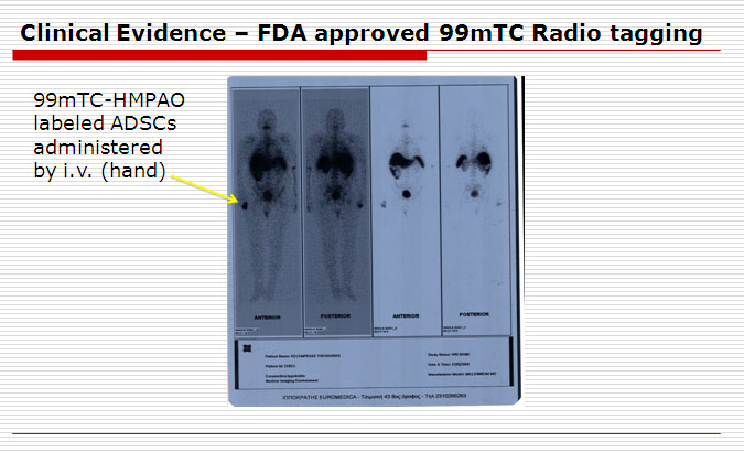 Of particular note to this scan was that the radio-tagged stem cells were found only on the right wrist, and not on the left, which clearly showed the ability of stem cells to go to the problem areas of the body.
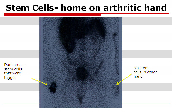 This is a closer view of the previously described scan which highlights the stem cells only on the right wrist.
EMPHYSEMA
When activated stem cells were administered via IV drip into the left arm, the stem cells went directly to the site of the inflammation in the lungs. Scan was taken 24 hours after stem cells were activated and radio tagged.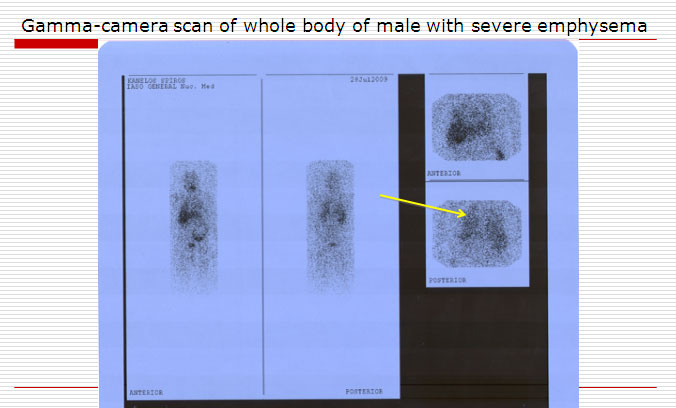 Fat-derived stem cells were again found on the affected area, which is the lung of an emphysema patient.
CEREBRAL PALSY
Below is an Indium scan of a 9-year old male diagnosed with Cerebral Palsy. The patient’s own adipose stem cells were harvested and activated. They were then tagged with Indium and returned the same day through an intravenous drip. A scan was taken and activated stem cells are clearly present in the brain on slide 15.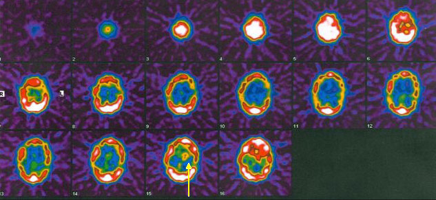 See Slide 15 above. Yellow-orange area indicates "hot spot" indium tagged stem cells. Below is a close up of slide 15. The yellow-orange color represents tagged stem cells that have homed on the area of the brain dysfunction.
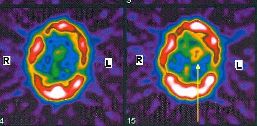 Slide 15: Yellow-orange area indicates indium tagged stem cells have logged in the affected brain tissue.
BRAIN INJURY
Particular case of a patient that had a brain injury. After IV infusion of patient's own fat-derived stem cells, scan clearly shows the stem cells on the affected area.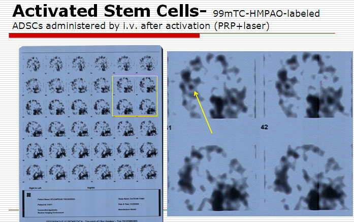
Recent Comments